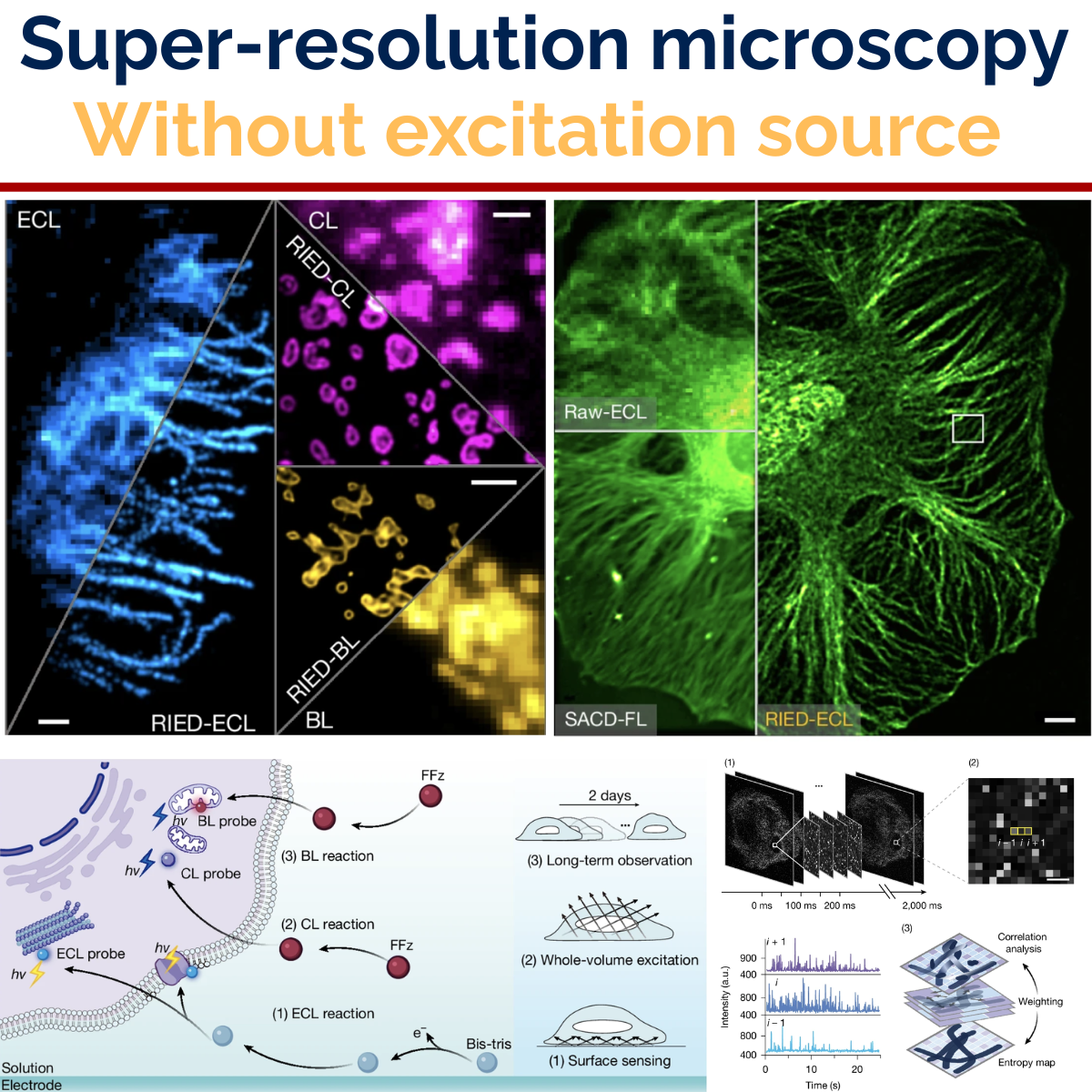
Every super-resolution technique most of us know — STORM, PALM, SIM, SOFI — starts the same way: you shine light on the sample to make it glow. It works beautifully, and it has reshaped what we can see inside cells. But that excitation light is also the source of two stubborn limits. It photobleaches the dye, and it slowly poisons the very cells you are trying to watch. There is always a clock running on a live-cell experiment.
A new paper in Nature removes the light source entirely. In Luminescent-reaction-enabled super-resolution imaging, Wenxin Zhu and colleagues in Jiandong Feng’s lab at Zhejiang University do super-resolution where the photons come from a chemical reaction happening in situ — not from a laser.
The old problem with light-free imaging
Reaction-excited luminescence is not new. Electrochemiluminescence (ECL), chemiluminescence (CL) and bioluminescence (BL) have all been used for imaging for years, and they are prized for one thing in particular: they emit against a truly dark background because nothing external is exciting the sample. Zero-background contrast, no phototoxicity ceiling.
The catch has always been that these reactions are dim. Conventionally you need seconds — sometimes tens of seconds — of exposure just to accumulate enough photons for a single frame. That is fatal for super-resolution, because most reconstruction methods depend on capturing fast, independent fluctuations. If one image takes half a minute, the fluctuations you need are long gone by the time you record them.
RIED: a reconstruction built for reaction photons
The authors’ answer is a framework they call RIED — reaction-enabled super-resolution imaging via entropy-weighted correlation combined with deconvolution. The important idea is that it was designed for the statistics of a chemical reaction, not borrowed from fluorophore-blinking methods.
RIED records the reaction as a spatiotemporal sequence, uses an entropy map to isolate genuine reaction “blinks” from background noise, correlates those events across space and time, and then applies a tailored sparse deconvolution to sharpen the result. When the authors ran the same data through conventional fluorescence super-resolution reconstructions, RIED outperformed them — precisely because it matches the underlying photon behaviour.
This is a different kind of contrast, not just a faster camera. In fluorescence, contrast comes from optical excitation. Here it comes from chemical reactivity — which means the image can, in principle, also carry functional information such as local variations in reaction kinetics.
A second, quieter contribution made the whole thing practical. On the ECL side, the team swapped the usual co-reactant (TPrA) for Bis-tris, a common biological buffer. That single change improved ECL cell-imaging brightness by about a thousandfold, letting them visualise microtubules at just 20 ms exposure — the difference between “too dim to resolve” and a workable super-resolution experiment.
What they actually resolved
The method holds up across all three luminescence chemistries, which is what makes it a general framework rather than a single trick.
What RIED demonstrated
- ~100 nm resolution in all three modalities — ECL reached 97 nm, CL 102 nm lateral, BL 117 nm.
- Full 3D ECL reconstruction resolving microtubules separated by 235 nm axially and 116 nm laterally.
- 8× lower detection threshold than fluorescence for the cancer biomarker CEA, helped by the absence of autofluorescence.
- 41 hours of continuous super-resolution live-cell imaging of mitochondria with no photobleaching — versus roughly 18 minutes before structured-illumination fluorescence bleached out.
- Mitochondrial transfer caught in action — that long observation window revealed two distinct modes of mitochondria moving between cells, dynamics that are essentially invisible to conventional imaging.
That 41-hour figure is the one I keep coming back to. It is not an incremental gain; it is a different regime. When your imaging is no longer bounded by how much light your sample can survive, you can watch slow biology unfold — and slow biology, like intercellular mitochondrial transfer, is exactly where a lot of interesting questions live.
Why this matters
It is tempting to file this under “yet another super-resolution method.” I think that undersells it. Most advances in the field push the same lever harder: brighter dyes, cleverer reconstruction, gentler illumination. RIED changes what the contrast is. The image is formed by chemistry rather than by optical excitation, which removes the phototoxicity ceiling instead of merely raising it.
There is a broader shift hiding in that sentence. For a century, image contrast in microscopy has been governed by excitation physics — what light you send in and how the sample scatters or re-emits it. Reaction-enabled super-resolution moves part of that control into chemistry: tune the reaction, and you tune the imaging. That opens a design space we have barely started to explore, and it is the kind of instrumentation-meets-chemistry frontier I find genuinely exciting.
Congratulations to the authors — Wenxin Zhu, Chi Zhang, Jiahui Gui, Yibo Yang, Yuxin Wan, Xin Wang, Liying Qu, Ao Guo, Ziqing Zhang, Zhenqian Han, Weisong Zhao and Jiandong Feng. You can read the full paper in Nature here.